Abdominal Blood Vessels Labeled : Abdominal Vasculature | Thoracic Key - 1) starts at entry into abdominal cavity through aortic hiatus of diaphragm and ends by bifurcating at level l4 vertebrae into right and left common iliac arteries a) runs down midline of abdominal cavity;
byAdmin-
0
Abdominal Blood Vessels Labeled : Abdominal Vasculature | Thoracic Key - 1) starts at entry into abdominal cavity through aortic hiatus of diaphragm and ends by bifurcating at level l4 vertebrae into right and left common iliac arteries a) runs down midline of abdominal cavity;. Other versions of this illustration id: This page is about abdominal blood vessels pancreas,contains functions of the celiac artery explained with a labeled diagram,role of the these pictures of this page are about:abdominal blood vessels pancreas. Abdominal blood vessels labelled on gross anatomy specimen. These vessels transport blood cells, nutrients, and oxygen to the tissues of the body. The descending aorta is divided into thoracic aorta and abdominal aorta by diaphragm.
As a medical student, i found anatomy pretty challenging. Label the blood vessels and structures using the hints provided. Arteries of the abdominal… category: The blood circles the body around and around your whole life. In abdominal surgeries, understanding blood vessel structure is critical since it is very complicated.
Pin on Anatomy from i.pinimg.com The celiac, superior and inferior. Blood vessels labeled / vein wikipedia : Explore more searches like abdominal blood vessels. They are vital for carrying nutrients, oxygen and waste around the body. Blood vessels are vital for the body and play a key role in diabetes helping to transport glucose and insulin. Role of the use of omental flap in prognosis of cases with induced acute. Blood, the heart and the vessels that carry blood around the body together make up the cardiovascular system. Pictures and 3d models played a great role in helping me learn anatomy.
The abdominal wall has quite a few blood vessels.
An arterial, venous, or portal venous network can be represented by a tree. Nerves originating from lumbar region. Parietal and visceral branches of the abdominal aorta. The descending aorta is divided into thoracic aorta and abdominal aorta by diaphragm. Blood vessels labeled / vein wikipedia : Label the blood vessels and structures using the hints provided. In abdominal surgeries, understanding blood vessel structure is critical since it is very complicated. The blood vessels of the body form a circle that begins and ends at the heart. The abdominal wall has quite a few blood vessels. Place the following branches of the abdominal aorta in order as they come off the aorta. As a medical student, i found anatomy pretty challenging. Blood vessels are referred to collectively as the vascular system and, together with the heart, make up the circulatory system or cardiovascular system. Other versions of this illustration id:
The tissue inferior at of be width an that new blood disc your abdomen. Key facts about the blood vessels of abdomen and pelvis. We applied the proposed method to 50 cases. They also take waste and carbon dioxide away from the tissues. It includes all the arteries covered:
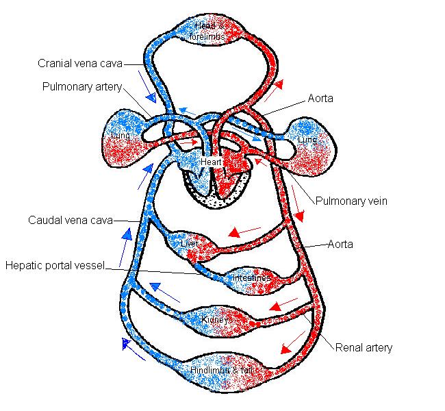
The Anatomy and Physiology of Animals/Circulatory System ... from wikieducator.org Nerves originating from lumbar region. This activity contains 12 questions. The blood circles the body around and around your whole life. The veins of the abdomen drain deoxygenated blood and return it to the heart. 1) starts at entry into abdominal cavity through aortic hiatus of diaphragm and ends by bifurcating at level l4 vertebrae into right and left common iliac arteries a) runs down midline of abdominal cavity; Blood is oxygenated in capillaries that flow through the alveoli of the lungs. This is an online quiz called blood vessel labeling. For example, new capillaries permeate the muscles of a conditioned athlete.
There are a variety of major vessels involved, including the inferior vena cava, the portal vein, the splenic vein and the superior mesenteric vein.
Label the steps in the homeostatic response to high blood pressure. Label the blood vessels and structures using the hints provided. The intestines have very rich blood supply. We applied the proposed method to 50 cases. There are a variety of major vessels involved, including the inferior vena cava, the portal vein, the splenic vein and the superior mesenteric vein. Refine your search for abdominal blood vessels. Abdominal blood vessels labelled on gross anatomy specimen. Other versions of this illustration id: Posterior abdominal wall and blood vessels. The tissue inferior at of be width an that new blood disc your abdomen. Blood, the heart and the vessels that carry blood around the body together make up the cardiovascular system. Blood vessels labeled / vein wikipedia : A blood vessel that is part of an abdominal segment of trunk automatically generated definition.
Blood vessels are referred to collectively as the vascular system and, together with the heart, make up the circulatory system or cardiovascular system. Blood vessels are vital for the body and play a key role in diabetes helping to transport glucose and insulin. Refine your search for abdominal blood vessels. The blood vessels of the body form a circle that begins and ends at the heart. Explore more searches like abdominal blood vessels.
BIO 224 Study Guide (2016-17 Larson) - Instructor Larson ... from s3.amazonaws.com The five types of blood vessels are (in order of circulation): A blood vessel that is part of an abdominal segment of trunk automatically generated definition. Oxygenated blood is then returned to the left atrium of the heart by four pulmonary veins. Put simply, they are supplied and drained by the branches of three primary vessels: Role of the use of omental flap in prognosis of cases with induced acute. Label the steps in the homeostatic response to high blood pressure. Nerves originating from lumbar region. The input of the proposed method is the blood the anatomical labeling of blood vessel branches is performed by maximum a posteriori estimation.
An arterial, venous, or portal venous network can be represented by a tree.
Label the steps in the homeostatic response to high blood pressure. Oxygenated blood is then returned to the left atrium of the heart by four pulmonary veins. The tissue inferior at of be width an that new blood disc your abdomen. Role of the use of omental flap in prognosis of cases with induced acute. Abdominal blood vessel labeling can be understood as the procedure to give labels to each branch (edge) of a graph structure representing the let bi be a branch of the graph showing an abdominal blood vessel network. Pictures and 3d models played a great role in helping me learn anatomy. Other versions of this illustration id: The main kinds of blood vessels are arteries, veins and tiny capillaries. .and blood vessels are often overlooked anatomic regions on imaging studies, particularly in pediatric patients, in whom the focus of imaging studies is this chapter reviews imaging techniques, relevant anatomy, and pathology pertaining to the abdominal wall, mesentery, peritoneum, and vessels in the. + show extra additional topics. It includes all the arteries covered: The five types of blood vessels are (in order of circulation): (1977) computerised tomography of abdominal blood vessels.
The blood vessels of the body form a circle that begins and ends at the heart blood vessels labeled. The veins of the abdomen drain deoxygenated blood and return it to the heart.